Rapid Profiling of Reduced and Intact Monoclonal Antibodies By LC/ESI-TOF MSRapid Profiling of Reduced and Intact Monoclonal Antibodies By LC/ESI-TOF MS

Figure 1.
As the pharmaceutical industry focuses on developing biotechnology-derived drugs, bioanalytical groups must produce generic methodologies for antibody characterization with higher throughput and faster sample turnaround times. Although antibody selectivity varies, overall antibody structures are highly conserved. Intact antibody LC/MS analysis is useful for profiling glycovariants and C-terminal Lys processing, and even more information can be gleaned from analysis of a reduced antibody.
UltraPerformance LC® (UPLC®) technology permits efficient desalting and rapid LC/MS mass profiling of an intact (4 min) and reduced (10 min) murine IgG1 monoclonal antibody. Highresolution mass spectrometry provides a powerful approach for profiling batch-to-batch variations and studying structural changes associated with drug production, formulation, and storage. These methodologies yield a simple, holistic view of the molecule and its variants by providing intact mass information that can be used to confirm primary structure and profiles of macroheterogeneity (glycosylation, Lys processing).
Results and Discussion
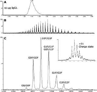
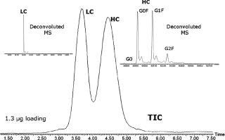
A fast (4-min cycle time) and efficient LC/ESI-MS method was used to profile multiple structural variants of an IgG. Figure 1 overlays TICs (y-axis linked) for this experiment and the associated summed mass spectra. The summed ESI-TOF mass spectrum for the antibody revealed a charge-state envelope over 2,300 to 4,000 m/z. The +51 charge state region has been enlarged (1c, inset) to show greater detail, indicating at least six significant antibody variants. The MaxEnt 1 deconvoluted spectrum clearly revealed carbohydrate-related heterogeneity. In Figure 2, rapid LC/ESI-MS was used to resolve and characterize the IgG heavy and light chains.
LC System: Waters ACQUITY UPLC® System
Column: Waters MassPREP™ Micro desalting column (2.1×5 mm)
Column Temperature: 80 °C
MS System: Waters LCT Premier™ ESI-TOF MS
Ionization Mode: ESI Positive, V mode
Capillary Voltage: 3,200 V
Cone Voltage: 40 V
Desolvation Temperature: 350 °C
Source Temperature: 150 °C
Desolvation Gas: 800 L/Hr
Ion Guide 1: 100 V (intact), 5 V (reduced)
Acquisition Range: 600 to 5,000 m/z
Sample: Monoclonal murine IgG1 (11.3 µg/µL, 0.1 M NaHCO3/0.5 M NaCl, ph 8.3
Mobile Phase: (A) 0.1% FA in H2O; (B) 0.1% FA in ACN
Gradient: 5-90% B in 2 min (intact); 10-50% B in 7.5 min (reduced)

Figure 1. ()

Figure 2. ()
You May Also Like


