North, South, East, and WestNorth, South, East, and West
February 1, 2014
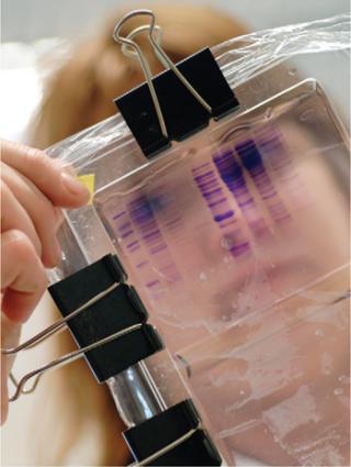
Electrophoresis is the basis of all blotting methods, and BPI Lab covered it last month (1). Electroblotting is a method for transferring electrophoretically separated proteins or nucleic acids onto a polyvinylidene fluoride (PVDF) or nitrocellulose membrane for permanence using electric current and a transfer buffer solution. This allows for analysts to further study them using probes, ligands, or stains. Capillary blotting is a variation designed to work with capillary electrophoresis.
After electrophoresis the following are stacked in cathode-to-anode order: a sponge, filter paper soaked in transfer buffer, the gel, a membrane, more soaked filter paper, and another sponge. When current is applied, it drives proteins or nucleic acids from the gel onto the membrane, where they are typically stained using a dye such as Coomassie Brilliant Blue to confirm that a sufficient quantity of material has been transferred. Immunoblotting involves the addition of specific antibodies with which proteins will interact. Affinity blotting involves other ligands in the same way.
A Brief History
The electroblotting technique was patented in 1989 by William J. Littlehales while he was working for American Bionetics (2). In 1991, BioGenex Laboratories acquired all the assets — including intellectual property — of that company when it went bankrupt. Before electroblotting, transferring gels to membranes was a more difficult and manual process.
Southern Blot: The names of all specific blotting methods come from the fact that the first was developed by Sir Edwin Southern, a British molecular biologist, when he was a professor at the University of Edinburgh in Scotland in the mid-1970s. Now a professor at Oxford, Southern is also chairman and chief scientific officer of Oxford Gene Technology. And he has been multiply awarded for his invention.
Northern Blot: Developers of the northern blot technique named it thus because of its similarity to the Southern technique, which came first. This variation came about in 1977 from the work of James Alwine, David Kemp, and George Stark at Stanford University.
Western Blot: The next method to appear originated almost simultaneously in the laboratory of Harry Towbin at the Friedrich Miescher Institute in Switzerland and the work of W. Neal Burnette, a postdoctoral researcher the Nowinski group at the Fred Hutchinson Cancer Center in Seattle. Burnette’s attempt at publication of his (slightly different) version of the technique was initially rejected — but his joking name for it caught on as colleagues and friends passed it around. The 1980s had arrived, and now the naming convention for blotting methods was set. In fact, when other scientists published a technique for DNA-binding proteins that combined Southern and western approaches in 1980, they named it the southwestern blot even though Burnette’s article hadn’t officially been published yet!
Eastern Blot: Further cementing the importance of Burnette’s work, multiple sets of authors have since dubbed a number of new methods eastern blotting. Among these is far-eastern blotting, which was developed (and named as such) in 1994 by scientists at the Tokyo Medical and Dental University in Japan.

A Sampling of Blotting Methods in the BPI Archives ()
Not surprisingly, with all the variations on the blotting theme, there are more suppliers providing services, membranes, reagents and probes, gels, instruments, and standards for this technology than can easily be counted. Among the most prominent are Abcam, Bio-Rad Laboratories, Cell Signaling Technology, Cyanagen, Li-Cor Inc., Life Technologies, Lonza, Molecular Devices, Molecular Probes, Perkin Elmer, Pierce Biotechnology, Protein Simple, Roche Applied Science, Rockland Immunochemicals, R&D Systems, sequence of a target gene’s protein product. Southern transfer also can be used to identify methylated sites in particular genes.
A Sampling of Blotting Methods in the BPI Archives
Mørtz E, et al. Proteomics Technology Applied to Upstream and Downstream Process Development of a Protein Vaccine. BioProcess Int. 6(1) 2008: 36–43.
Aschermann K, Lutter P, Wattenberg A. Current Status of Protein Quantification Technologies. BioProcess Int. 6(4) 2008: 44–53.
Mücke M, Ostendorp R, Leonhartsberger S. E. coli Secretion Technologies Enable Production of High Yields of Active Human Antibody Fragments. BioProcess Int. 7(8) 2009: 40–47.
Wang X, et al. Improved HCP Quantitation By Minimizing Antibody Cross-Reactivity to Target Proteins. BioProcess Int. 8(1) 2010: 18–24.
Liu X, et al. Isolation of Novel High-Osmolarity Resistant CHO DG44 Cells After Suspension of DNA Mismatch Repair. BioProcess Int. 8(4) 2010: 68–76.
Schwertner D, Kirchner M. Are Generic HCP Assays Outdated? BioProcess Int. 8(5) 2010: 56–62.
Hoffman K. Letter to the Editor. BioProcess Int. 8(11) 2010: 8–10.
Menendez AT, et al. Recommendations for Cell Banks Used in GXP Assays. BioProcess Int. 10(1) 2012: 26–40.
Liu N, et al. Host Cellular Protein Quantification. BioProcess Int. 10(2) 2012: 44–50.
Ausubel LJ, et al. Production of CGMP-Grade Lentiviral Vectors. BioProcess Int. 10(2) 2012: 32–43.
Murray EW, et al. A Host Cell Protein Assay for Biologics Expressed in Plants. BioProcess Int. 10(9) 2012: 44–51.
Cossins A, Hooker A. Preformulation Development of a Recombinant Targeted Secretion Inhibitor. BioProcess Int. 11(2) 2013: 52–57.
Mire-Sluis A, et al. Drug Products for Biological Medicines. BioProcess Int. 11(4) 2013: 48–62.
Detmers F, Mueller F, Rohde J. Increasing Purity and Yield in Biosimilar Production. BioProcess Int. 11(6) 2013: S36–S41.
Berkelman T, et al. Enhanced 2-D Electrophoresis and Western Blotting Workflow for Reliable Evaluations of Anti-HCP Antibodies. BioProcess Int. 11(8) 2013: 50–61.
Gupta D, Prashanth GN, Lodha S. A CMO Perspective on Quality Challenges for Biopharmaceuticals. BioProcess Int. 11(9) 2013: 20–26.
The northern blot is used in molecular biology research to study gene expression by detection of RNA (or isolated mRNA). Analysts can observe a particular gene’s expression pattern among tissues, organs, developmental stages, environmental stress levels, pathogen infection, and over the course of treatment. So this technique is used in diagnostics as well as cell-line analysis. Online databases help scientists compare their results to published examples. Although gene expression can be analyzed using many different methods (e.g., polymerase chain reaction, RNase protection assays, and microarrays), northern blotting can detect small changes in gene expression that other methods may not. However, it is limited in scope, usually involving one or a small number of genes at a time — but membranes can be stored and used for further analysis years later.
Western Blot: Sometimes called protein immunoblotting, the western technique is widel
y accepted for detecting specific proteins in complex samples. After sodium-dodecyl sulfate polyacrylamide gel electrophoresis (SDS-PAGE) (1), separated proteins are transferred to a membrane and stained with antibodies specific to a target protein. With applications in diagnostics and protein expression analysis (among others), this technique is very widely used in biopharmaceutical laboratories. Excellent western blotting guides can be found online from suppliers such as Abcam plc, Bio-Rad Laboratories, and GE Healthcare (4,5,6). And the Western Blot Encyclopedia from Sino Biological Inc. is another valuable online resource (7).
Eastern Blot: Although many definitions have been applied to the name, in general eastern blotting is used to analyze posttranslational modifications of proteins (often carbohydrate epitopes in glycosylation). This makes it useful for sponsors of complex protein products.
About the Author
Author Details
Cheryl Scott is cofounder and senior technical editor of BioProcess International, 1574 Coburg Road #242, Eugene, OR 97401; 1-646-957-8879; [email protected].
REFERENCES
1.) Scott, C 2014. Analysis By Size and Charge: SDS-PAGE, Capillary, and Isoelectric Focusing Techniques Anchor the Biopharmaceutical Laboratory. BioProcess Inc. 12:26-29, 49.
2.) Littlehales, WJ.
3.) Bowen, B. 1980. The Detection of DNA-Binding Proteins By Protein Blotting. Nucl. Acids Res. 8:1-20.
4.).
5.).
6.).
7.).
You May Also Like


